Mental growth of child into adult

Licensed Mental Health Counselor

Maslow’s Hierarchy of Needs is based on Dr. Abraham Mallow’s research and hypothesis. It describes the stages we all need in order to become fully functioning and responsible adults moving towards reaching the highest possible achievements humans can accomplish.

Maslow's_hierarchy_of_needs

The hierarchy is broken down into five needs:

  • Self-Actualization           
  • Esteem
  • Love/Belonging
  • Safety
  • Physiological

The physiological needs is all the basic needs someone needs to survive such as food, water, air, homeostasis, excretion, and health.

The safety and security needs include security of:  body, resources, morality, family, health, stability and protection. At this level, all of the child’s physiological needs have been taking care of and they are interested in finding safety, such as from strangers. At this stage, a child develops a need for limits, order and structure. This is also the stage were fears and worries develop. The child may start fearing the dark, strange noises in their bedroom, or being kidnapped.

At the love and…

View original post 1,201 more words

Hydrocephalus & Shunts: what the Neurologist should know

HYDROCEPHALUS AND SHUNTS: WHAT THE NEUROLOGIST SHOULD KNOW (by Dr. Vaseem Zamair)

The second most common reason for being sued for negligence in neurosurgery is a problem related to hydrocephalus management (the first being spinal surgery!). However, the good news is that the overall standard of care for patients with hydrocephalus appears to have greatly improved over the last 10 years with the advent of better facilities for investigation, new approaches to treatment, and a greater awareness of the need for adequate follow up. In the possible absence of a local neurosurgeon with an interest in hydrocephalus, a neurologist who is faced with the ongoing care of a patient with hydrocephalus should ideally have a clear idea of what exactly constitutes appropriate follow up and which clinical and radiological warning signals of shunt problems to look out for.

DEFINITIONS

Hydrocephalus is an excessive accumulation of cerebrospinal fluid (CSF) within the head caused by a disturbance of formation, flow or absorption.

“Hydrocephalus ex vaccuo” is a misnomer. It refers to asymptomatic ventricular enlargement caused by generalised loss of cerebral tissue, from severe head injury, infarction or cerebral hypoxia.

BASIC HYDROCEPHALUS PATHOPHYSIOLOGY IN ADULTS

The normal CSF production rate in an adult is 0.35 ml/min (20 ml/hour or 500 ml/24 hours). The capacity of normal lateral and third ventricles is approximately 20 ml, whereas the total CSF volume in an adult is 120–150 ml. Hence, in normal circumstances CSF is recycled over three times each day.

ICP rises if production of CSF exceeds absorption, but CSF production will fall as ICP rises to high levels, and compensation (stabilisation of hydrocephalus at a new steady state) may occur through transventricular absorption of CSF. Dural absorption may also be important through nerve root sleeves and unrepaired meningocoeles. Of vital significance is the fact that compensated hydrocephalus (for example, in patients with a longstanding non-functional shunt) is not necessarily permanent because of the sometimes precarious nature of the balance between production and absorption of CSF. Clinicians involved in the care of patients with so called stable hydrocephalus should always be alert to the possibility of insidious subclinical progression or late decompensation of this condition, that may occur spontaneously or after a minor head injury.

As hydrocephalus develops the temporal and frontal horns dilate first, often asymmetrically. There is then elevation of the corpus callosum and stretching of the white matter tracts followed by thinning of the convexity grey matter of the brain

CLINICAL FEATURES

The clinical features of hydrocephalus are notoriously variable, depending on the rapidity of onset of the condition (see box). The most rapid deteriorations are seen in young adults with colloid cysts of the third ventricle where the acute rise in ICP caused by the ball valve plugging of the third ventricle can lead to sudden death. The least rapid presentations occur in older patients with soft compliant brains where the only clue to the presence of progressive hydrocephalus may be a subtle slowing of gait or mentation. This slow presentation also characteristically occurs after severe head injury or subarachnoid haemorrhage.

INVESTIGATIONS

Computerised tomography

A computerised tomographic (CT) scan should be undertaken to assess the overall size of the ventricles, and to determine if periventricular oedema or “lucency” is present. A CT scan is also useful to assess the size of the fourth ventricle—if large, this suggests a communicating hydrocephalus, whereas a relatively small fourth ventricle implies obstructive hydrocephalus that might be best treated by endoscopic third ventriculostomy rather than a ventriculo-peritoneal shunt.

Magnetic resonance imaging

Chiari malformations and cerebellar or periaqueductal tumours, sometimes not visible on CT, can be detected with magnetic resonance imaging (MRI). This imaging technique is also useful for detecting flow voids in the third ventricle and aqueduct. Various radiological features (angle of the frontal horns on coronal view, downward bowing of the floor of the third ventricle, elevation of the corpus callosum) may imply active hydrocephalus, but these must not be heavily relied upon because of their poor sensitivity. Differentiating normal pressure hydrocephalus from cerebral atrophy is still often more of an art than a science.

ICP monitoring/CSF infusion studies

ICP monitoring and CSF infusion studies are now being used more frequently in young patients with mild symptoms and older patients with possible low grade hydrocephalus. ICP monitoring may reveal “B waves” either at night time alone or throughout the day and night. An ICP above 15 mm Hg at frequent intervals during the night or day while asleep or resting is abnormal, and patients with functioning shunts should normally have an ICP below or near to zero while 45° head up in bed.

Clinical features of hydrocephalus

  • Young adults:
    • Symptoms—headache, vomiting, failing vision, drowsiness, “muzziness of the head”, fatigue
    • Signs—papilloedema, enlarged blind spots on visual field analysis or reduced visual acuity, failure of upward gaze, general clumsiness, dyspraxic gait, large head
  • Older adults/elderly:
    • Symptoms—slowing of mental capacity, unsteady on feet/frequent falls, incontinence, drowsiness, headaches less frequently
    • Signs—gait dyspraxia (slow, hesitant shuffling gait), dementia (reduced mini-mental score), rarely papilloedema

Lumbar CSF infusion tests measure CSF outflow resistance, which in simple terms represents the overall compliance of the intracranial and spinal CSF compartment. During this test saline or artificial CSF is constantly infused via a lumbar puncture needle or catheter, and the subsequent gradient of rise in the ICP with time is recorded. A low outflow resistance corresponds to high cerebral compliance and vice versa. Normal values are 5–10 mm Hg/ml/minute and a value > 18 mm Hg/ml/minute appears to be the approximate cut off point for diagnosing active hydrocephalus in the elderly. Other compliance monitors have recently been developed that are placed as bolts through small twist drill holes in the skull. These tests can be used to guide treatment of patients with newly diagnosed ventricular enlargement; they can also be useful in patients with possible blockage of their shunts or delayed occlusion of their third ventriculostomy site.

Transcranial Doppler

Transcranial Doppler involves the non-invasive measurement of the middle cerebral artery flow velocities and pulsatility index. The latter index appears to correlate fairly well with ventricular distension and cerebrovascular impedance. The quality of information obtained is very much operator dependent, but can be useful for monitoring patients in an outpatient setting.

Tympanic membrane displacement

Measurement of tympanic membrane displacement is an indirect non-invasive technique for assessing ICP. It is based on a direct communication of pressure waves from the intracranial space to the middle ear via the endolymphatic system of the inner ear. There must be a clear ear canal, intact tympanic membrane, and skilled, experienced operators to give dependable results. In order to diagnose a blocked shunt the patient should preferably already have a baseline reading while well with an obviously functioning shunt.

CSF sample

In post-subarachnoid and post-meningitic hydrocephalus, CSF samples are useful for cell counts, protein concentration, and to exclude residual infection. A protein concentration greater than 4 g/l will clog up most ventriculo-peritoneal shunt valves.

Psychometric analysis

Although it is highly unlikely that any patients with possible pressure from hydrocephalus would ever stand a chance of seeing a neuropsychologist during an investigational work up, the right posterior hemisphere has been shown to be most susceptible to functional deterioration from raised ICP. Deterioration in hand–eye coordination and visuospatial skills may precede classic symptoms of shunt blockage.

WHICH TREATMENT OF HYDROCEPHALUS IS BEST: ENDOSCOPIC OR SHUNT?

Since the 1960s the standard treatment of hydrocephalus has been the insertion of a valved ventricular shunt either into the peritoneum or right atrium of the heart. Most neurosurgeons have used ventriculo-peritoneal (VP) shunts mainly because of the potentially life threatening nature of some of the complications of ventriculo-atrial (VA) shunts (multiple pulmonary emboli, shunt nephritis).

Over the past 10 years or so there has been a huge resurgence of interest in ways of avoiding CSF shunts for hydrocephalus by utilising endoscopic techniques, such as third ventriculostomy. During this period smaller endoscopes have been developed that have better optics and brighter light sources, and these changes have undoubtedly enabled neurosurgeons to perform these techniques with a greater degree of confidence and safety for the patient. The attraction of giving patients the chance of escaping the numerous inherent complications of CSF shunts has led to a rapid expansion of enthusiastic neurosurgeons offering a neuroendoscopic alternative for the treatment of hydrocephalus. Techniques such as endoscopic septostomy (making a hole in the septum pellucidum for a trapped ventricle), cyst fenestration or choroid plexus coagulation have been used with reasonable long term success, and colloid cysts and pineal tumours are often ideally treated by skilled neuroendoscopy.

ENDOSCOPIC THIRD VENTRICULOSTOMY

In the past the main indication for third ventriculostomy has been non-communicating (obstructive) hydrocephalus, demonstrated by CT or ventriculography. There would typically be dilatation of the lateral ventricles, ballooning of the third ventricle, and a relatively small fourth ventricle. The aetiology of hydrocephalus was generally congenital aqueduct stenosis, and a history of meningitis would have been regarded a contraindication. However, these traditional criteria are no longer adhered to by an increasing number of neuroendoscopists, who have greatly extended their indications for third ventriculostomy to include patient groups previously regarded as inappropriate.

The technique consists of the creation of a single burrhole in the frontal region followed by ventricular cannulation and insertion of a 3 or 4 mm wide neuroendoscope into the lateral ventricle. The third ventricle is then negotiated via the foramen of Monro and a hole is then made in the floor of the third ventricle between the infundibulum of the pituitary gland and the mammillary bodies (fig 2). This creates a CSF fistula between the third ventricle and the subarachnoid space in front of the brainstem. The hole is made with a small electrode and enlarged with a balloon dilator, such that a very exciting close up view of the basilar artery is unveiled.

The cause of hydrocephalus undoubtably influences the chance of long term success. There is clearly a better success rate in patients with aqueduct stenosis, spina bifida, and tectal, pineal, and posterior fossa tumours. The risk of failure appears to increase with a past or recent history of intracranial infection, presumably as a result of obliteration of cerebrospinal fluid pathways. Long term results have been questioned as follow up is often not more than five years in the literature, but most studies quote an overall success rate between 65–75%.

The overall complication rates reported in published series varies widely from 4–30%, but the overall rate of serious complications is 9.4% and this includes an average 3% infection rate, 2.3% haemorrhage rate, and 1.3% risk of a permanent neurological deficit. Fortunately the rate of life threatening complications appears to be low and postoperative deaths are rare (0.1%, or 1 per 1000 third ventriculostomies).

CEREBROSPINAL FLUID SHUNTS

Most shunting systems drain according to the differential pressure gradient between the ventricle and the tip of the distal catheter. These valves have been shown to be effective in the majority of patients and a typical valve is shown in fig 3. Most neurosurgeons use medium pressure valves, that will drain CSF continuously if the differential pressure is over about 10 mm Hg. The ventricular catheter of a shunt is normally inserted through a burrhole in the right parieto-occipital region and the valve will sit usually behind the right ear. The distal catheter is tunneled subcutaneously down to another incision in the abdomen where it is then placed into the peritoneal cavity. It is not usually helpful for non-neurosurgeons to palpate or flush the shunt valve, as their contours and characteristics are so variable as to make interpretation notoriously inaccurate.

Clinical features of shunt malfunction

  • Headaches
  • Vomiting
  • Drowsiness
  • Papilloedema with or without failing vision
  • Occasionally failure of upward gaze
  • Neck stiffness
  • Thoracic back pain in patients with spina bifida
  • CT scan (enlargement of ventricles)
  • Plain x ray of shunt system (lateral skull, anteroposterior (AP) chest and AP abdomen)
  • Palpation of shunt reservoir—unreliable
  • Peripheral blood for C reactive protein, white cell count if there has been any recent surgery
  • Shunt reservoir tap
  • ICP monitoring/lumbar infusion test

Investigations of shunt malfunction

In selected patients a ventricular access device (otherwise known as an “Ommaya reservoir”) is placed in the right frontal region for ICP monitoring or treatment of infections (fig 4). These cannot be flushed or assessed in any way by palpation, but provide the facility for potentially life saving percutaneous aspiration of CSF in the event of acutely raised ICP. This can be done by any clinician in this situation and simply involves the passing of a butterfly needle through the skin perpendicular to the surface of the skin at the apex of the dome of the reservoir, until a “pop” is felt. Elective sampling of reservoirs or shunts should preferably be carried out by a neurosurgeon, unless the clinician looking after the patient has had previous experience in the technique.

Differential pressure valves allow the siphoning effect in the upright position and this may lead to excessive CSF drainage from the ventricles. Some systems now incorporate an anti-siphon device to ameliorate this effect.

“Programmable” or adjustable valves allow the closing pressure to be altered externally using a special magnetic adjusting device. Although this is sometimes extremely useful in selected shunted patients with intractable headaches, it can lead to problems following inadvertent change of pressure—for example, by having an MRI scan or, less obviously, by using headphones and certain cordless phones.

Flow controlled valves, such as the Orbis-Sigma valve, have a more physiological CSF drainage pattern, but they do not appear to be effective in normal pressure hydrocephalus or where brain compliance is poor (so called “brittle ventricles”).

COMPLICATIONS OF SHUNTING

Shunt obstruction

Shunt obstruction may occur proximally in the ventricular catheter as a result of choroid plexus, red cells, tumour cells, or a high protein concentration in the CSF. Blockage of the distal catheter can occur as a result of body growth (if the shunt was placed during childhood), adhesions within the abdominal cavity, especially when associated with a low grade infection, pregnancy, and occasionally constipation.

Urgent help from the on-call neurosurgeon should be sought for all suspected cases of acute shunt malfunction as patients with little remaining compensatory reserve may deteriorate suddenly as a result of a respiratory arrest, seizures, or simple coning. Shunt blockage may cause death and blindness if there is a combination of sudden onset and delay in treatment.

Uncomplicated shunt revisions do not affect long term outcome. However, revision surgery on patients with blocked shunts is occasionally complicated by serious secondary ventricular or intraparenchymal haemorrhage, and any patient who is “not quite right” soon after a shunt revision should have a follow up CT scan immediately. Most young patients with shunts require a revision operation once or twice every 10 years as the shunt tubing degenerates gradually over the years and flakes of silicone break off (causing subcutaneous granulomas), weakening the wall of the tube. Eventually the tube may fracture or obstruct.

Some unlucky patients run into multiple problems with their shunt, usually as a result of “one problem leading to another” (fig 5). There is little to be done in such circumstances, other than commiserate with the patient and approach each hurdle in a positive and objective manner. However, when the situation becomes unduly complicated or intractable it may be prudent for the neurologist or neurosurgeon to seek the opinion of a neurosurgeon with a special interest in hydrocephalus.

Infection

Shunt infections are usually caused by the patient’s own skin organisms (most common is Staphylococcus epidermidis), which gain access to shunt tubing during the shunt procedure. Typically this contamination will cause an internal shunt colonisation where the bacteria settle and grow on the internal wall of the shunt catheter and valve, establishing adherent colonies. However, some bacteria set up a ventriculitis without full colonisation of the shunt, and others (for example, Staphylococcus aureus) cause an external shunt infection (deep wound infection).

The most important clinical features of a shunt infection are as follows:

  • general malaise
  • pyrexia
  • headaches, vomiting, neck stiffness
  • abdominal tenderness or distension
  • recurrent lower end shunt obstruction
  • occasionally pain and erythema around the shunt
  • pulmonary hypertension or shunt nephritis in chronic VA shunt infections
  • recent shunt operation
  • 90% of VP shunt infections present within three months of a shunt operation
  • raised C reactive protein
  • high peripheral and CSF white cell count
  • culture of organism from CSF.

Patients should be reassessed urgently by the relevant neurosurgical team should any of the above symptoms develop within the first few months after a shunt operation. After six months a VP shunt will not become infected unless intra-abdominal sepsis occurs (for example, appendicitis, diverticulitis or postgastrostomy feeding tube insertion).

The current incidence of shunt infection in most neurosurgical units is about 5–8%, but many units are now achieving better results as a result of preventative measures and protocols. Details, including infection rates, of most shunt operations performed in the UK are now submitted to the UK Shunt Registry that was set up by the Medical Devices Agency a few years ago. It is hoped that information derived by analysing this huge data set will lead to further improvements in the standards of shunt surgery in the future.

Infected shunts will often have to be removed and then replaced after two weeks of antibiotics and temporary drainage. Infections with low grade pathogens can sometimes be treated with intraventricular and intravenous antibiotics alone.

Over-drainage

The perfect shunt valve has yet to be designed and many current models allow over-drainage of CSF due to the siphoning effect. The hydrostatic pressure (25–75 cm CSF) caused by the weight of the column of CSF within the distal catheter leads to fluid being sucked out of the ventricles in the upright position. The valve pressure may be set too low for an individual patient leading to over-drainage; this can be remedied by adjustments of the valve pressure either by revision of the valve or by using a programmable valve. ICP monitoring may be required in those patients presenting with possible low pressure headaches without evidence of subdurals on the CT scan.

Subdural haematoma can occur during the first six months after a shunt insertion and has been shown to be related to the amount of CSF released at operation. Small collections occur in up to 30% of patients after shunt insertion in the elderly, but symptomatic collections requiring surgery affect only 10–15%. The symptoms of a shunt related subdural collection include headaches, confusion, hemiparesis, and drowsiness.

FOLLOW UP AFTER ENDOSCOPIC THIRD VENTRICULOSTOMY

There is a clear need for continued follow up of patients after apparently initially successful third ventriculostomy, as late failure occurs in up to 40% and sudden death has been recently reported as little as two years following apparently successful third ventriculostomy.

Most patients are followed up primarily by monitoring their clinical symptoms, optic fundi, and visual acuity. Midline sagittal T2 weighted MRI sequences combined with cine phase contrast MRI flow measurements provide a reliable tool for ascertaining the patency of the stoma during follow up evaluation. The important sign is flow voids through the ostomy; change in ventricular size is less important after third ventriculostomy. However, a 33% reduction in the size of the third ventricle on follow up CT scans occurs in patients whose symptoms have been successfully controlled. Lateral ventricles will only decrease in size on average by 16%.

ICP measurements from reservoirs may be useful in selected younger patients, as the value of ICP monitoring in shunted children by means of a reservoir or by parenchymal devices has been previously well documented. Reservoirs with an integral telesensor to measure ICP non-invasively may prove useful after third ventriculostomy where the preoperative pressures have been substantially raised. Other workers have advocated the use of CSF infusion studies to monitor cerebral compliance, or transcranial Doppler indices before and after operation as an indirect measure of compliance.

HOW SHOULD WE FOLLOW UP SHUNTED PATIENTS?

Arrangements for follow up of adult patients with shunts have been extremely variable, with some patients receiving careful regular annual outpatient assessments and others being completely discharged and left to contact their general practitioners in the event of symptoms. A number of patients have gone blind or died as a result of undetected chronic shunt malfunction, and efforts by the Association of Spina Bifida and Hydrocephalus have led to improvements in standards of follow up care. The principle components of good long term care of patients with shunted hydrocephalus are outlined below.

All patients should have a baseline CT brain scan 6–12 months after their initial shunt insertion, while they are well. They should preferably retain a copy of this baseline scan if they travel far from their base neurosurgical unit, and it is our experience that most patients are willing to pay for this copy. Although it is not always possible to detect shunt blockage on a CT scan as a result of a patient having non-compliant ventricles, this is the exception rather than the rule.

All patients and carers should be given clear instructions (preferably written) as to what symptoms to look out for and when to contact their doctor. Some documentation of exactly which type of valve has been implanted should be given to the patient. Those with programmable/adjustable valves should always have their shunt re-programmed, or at least checked by their neurosurgeon after any MRI scan has been carried out. They should be made aware of the possible problems of inadvertent valve pressure change from extraneous magnetic sources.

All younger (under 60 years) patients should have an annual visual acuity check by an optician or an ophthalmologist if there have been particular concerns about vision.

All younger patients with a shunt should probably be encouraged to seek a neurosurgical check up at least every three years, ideally at a dedicated hydrocephalus follow up clinic.

Tagged , , , , , , , , , , , , , , , , , ,

Basics of Dermatology & Gonorrhoeae Infection

 Dermatology & Flora  (by Dr. Vaseem Zamair)


 Image

Epithelium – made of  Keratinized, Stratified, Squamous cells

Every cell has 3 sides: APICAL – upper, LATERAL –side, BASAL –inner or lower part.

Apical domains have cilia, villus and microvilli on them

Lateral Domains have different type of junctions which help either to attach to other cell or not to let anything pass thru it

  1. Zona occludens – tight junction which doesn’t let any thing to pass thru it – Virus and bacteria damaging zona occludens are Clostridium and Helicobacter Pylori.
  2. Zona Adherence – intermediate junction
  3. Macula adherence – Stick buttons like dics – which are called desmosomes. Antibodies against its own desmosomes make Pemphigus Vulgaris  –à multiple blisters form
  4. Gap Junctions – it is the real connection between 2 adjacent cells where cytoplasms are interconnected… but this Gap-junctions closes if there is increase in Calcium or Potassium

Basal Domain – it has

Zona Lucida – Integrins, lemanins, Enactins

Zona Densa  – collagen 4, Fibronectin                   both make Basal lamina

Lamina reticularis – collagen 7 and reticularis with collagen 3

External lamina


Common Inflamations

Image

Cellulitis – diffuse inflammation of connective tissue with severe inflammation of dermal and subcutaneous layers of the skin.

Cellulitis can be caused by normal skin flora or by exogenous bacteria, and often occurs where the skin has previously been broken: cracks in the skin, cuts, blisters, burns, insect bites, surgical wounds, or sites of intravenous catheter insertion (Red, Warmth and Pain).

Image

Panniculitis – involve fat around the inflamation.

Image

Oomphalitis – surround the belly button ( in new borns).

Image

Carbuncle – when it is boil, firm and indurated.

Image

Furuncle – if hair follicle is involved.

 Image

Folliculitis – if pus at base of hair follicle & it proliferates to other hair follicles.

Image

Fasciitis – when fascia is involved (pain during movement) *

Skin and Mucosal surfaces most common NORMAL FLORA


Aerobic = who are exposed to Oxygen (most of Gram+)

Anaerobic = who hide from Oxygen (can be both)

Gram- (negative)  = who dwell in places which are not exposed to high concentration of Oxygen (can be called facultative too).

SKIN ( mostly aerobic and gram+ )


  1. Staph. Aureus   à most common in all skin diseases & rash
  1. Staph. Epidermitis à present in deep surfaces of skin ( deep skin infections, I.V. lines, shunts passed etc)

3. Strep. Pyogenes à special types of rash ie.

a. Honey crusted lesions – Impetigo

b. Sand paper rash which includes palm & soles with Strawbery tongue – Scarlet fever

c. Necrotizing fasciitis – deep fascia is involved

d. Lymphangitis – RED STREAK on lymphatic chanel area (since strep. Travels thru lymph chanels)

e.  Erysepelas – infection of SUB-CUTANEOUS FAT, doesnot blanch and is with raised borders of rash.

4. Propionobact. Acne à gram+ and anaerobic, dwelling in hair follicle loves oil – propionic acid. Therefore increase of progesterone levels always cause acne (during teenage years in Males and every month is females).

5. Staph. Saprophyticus à most commonly present in females and causes urinary tract infection as easily ascend through vagina.

4. and 5.  hide from Oxygen à Anerobic (produce GAS *)

MUCOSAL SURFACES

I. Nose – Staph. Aureus

II. Throat – Strep. Pyogenes

III. back of Throat :

  1. Strep. Pneumococcus: gram+
  2. Hemophi. Influenza
  3. Nisseria all types

IV. Beneath Gums and teeth : hide from Oxygen, therefore ANEROBIC àproduce Gas*

    1. Peptostreptococcus
    2. Actinomyces
    3. Fusobacterium
    4. Strep. Viridans
    5. strep. Mutans
    6. strep. Sangris
    7. strep. Salivaris

NEISSERIA GONORRHOEAE


Image

 often called the gonococcus, causes the second most commonly transmitted sexual disease, gonorrhea (chlamydial infections are slightly more common).

Virulence factors of the gonococcus include:

1) Pili: Neisseria gonorrhoeae has complex genes coding for their pili. These genes undergo multiple recombinations, resulting in the production of pili with hypervariable amino acid sequences. These changing antigens in the pili protect the bacteria from our antibodies, as well as from vaccines aimed at producing antibodies directed against the pili

2) Protein II: This outer membrane protein is also involved in adherence to host cells.


Image

Gonococcal Disease in Men

A man who has unprotected sex with an infected person can acquire a Neisseria gonorrhoeae infection. This organism penetrates the mucous membranes of the urethra, causing inflammation of the urethra (urethritis). Although some men will remain asymptomatic, most will complain of painful urination along with a purulent urethral discharge (pus can be expressed from the tip of the penis). Both asymptomatic and symptomatic men can pass this infection to another sexual partner. Possible complications of this infection include epididymitis, prostatitis, and urethral strictures. Fortunately, this disease is easily cured by a small dose of ceftriaxone.

Image

Gonococcal Disease in Women

Like men, women can also develop a gonococcal urethritis, with painful burning on urination and purulent discharge from the urethra. However, urethritis in women is more likely to be asymptomatic with minimal urethral discharge. Neisseria gonorrhoeae also infects the columnar epithelium of the cervix, which becomes reddened and friable, with a purulent exudate. A large percentage of women are asymptomatic. If symptoms do develop, the woman may complain of lower abdominal discomfort, pain with sexual intercourse (dyspareunia), and a purulent vaginal discharge. Both asymptomatic and symptomatic women can transmit this infection. A gonococcal infection of the cervix can progress to pelvic inflammatory disease (PID, or “pus in dere”). PID is an infection of the uterus (endometritis), fallopian tubes (salpingitis), and/or ovaries (oophoritis).

Clinically, patients can present with fever, lower abdominal pain, abnormal menstrual bleeding, and cervical motion tenderness (pain when the cervix is moved by the doctor’s examining finger). Menstruation allows the bacteria to spread from the cervix to the upper genital tract. It is therefore not surprising that over 50% of cases of PID occur within one week of the onset of menstruation. The presence of an intrauterine device (IUD) increases the risk of a cervical gonococcal infection progressing to PID. Chlamydia

trachomatis is the other major cause of PID

Complications of PID include:

1) Sterility: The risk of sterility appears to increase with each gonorrhea infection. Sterility is most commonly caused by scarring of the fallopian tubes, whichoccludes the lumen and prevents sperm from reaching the ovulated egg.

2) Ectopic pregnancy: The risk of a fetus developing at a site other than the uterus is significantly increased with previous fallopian tube inflammation (salpingitis). The fallopian tubes are the most common site for an ectopic pregnancy. Again, with scarring down of the fallopian tubes, there is resistance to normal egg transit down the tubes.

3) Abscesses may develop in the fallopian tubes, ovaries, or peritoneum.

4) Peritonitis: Bacteria may spread from ovaries and fallopian tubes to infect the peritoneal fluid .

5) Peri-hepatitis (Fitz-Hugh-Curtis syndrome): This is an infection by Neisseria gonorrhoeae of the capsule that surrounds the liver. A patient will complain of right upper quadrant pain and tenderness. This syndrome may also follow chlamydial pelvic inflammatory disease.

Diagnosis and Treatment:

Diagnosis of Neisseria gonorrhoeae infection is best made by Gram stain and culture on Thayer-Martin VCN medium. Pus can be removed from the urethra by inserting a thin sterile swab. When this is Gram stained and examined under the microscope, the tiny dough-nut shaped diplococci can be seen within the white blood cells. In the past, the combination of penicillin G with probenicid was the regimen of choice. However, there arose penicillinase-producing gonococcal strains and now an even tougher strain, with chromosomally mediated antibiotic resistance to many antibiotics, such as tetracycline, erythromycin, and trimethoprim) sulfamethoxazole. This resistance is mediated by a block in antibiotic penetration into the bacterial cell. The current therapy of choice is ceftriaxone, a third generation cephalosporin. Ceftriaxone will also treat syphilis. If the patient is allergic to cephalosporins, spectinomycin or ciprofloxacin can be used as an alternative. The patient should also be treated at the same time with doxycycline or azithromycin for Chlamydia trachomatis, because up to 50% of patients will be concurrently infected with this beta-lactam-resistant (ceftriaxone included) bacteria.

Tagged , , , , , , , , , , , ,

PHEOCHROMOCYTOMA – a short picture notes

PHEOCHROMOCYTOMA  (by Dr. Vaseem Zamair)

It is not adrenal gland tumor!!! But it is a tumor of Neuroendocrine cells called CHROMAFFIN CELLS present in medulla of adrenal gland (80%) and other ganglia of sympathetic nervous system,

Image

and they secrete adrenaline (epinephrine), noradrenaline (norepinephrine), and enkephalin and enkephalin-containing peptides into the blood stream and to adjacent organs or tissue to produce sympathetic nervous system hyperactivity, where BP is persistent even after drug therapy.

Image

Symptoms: Head ache, sweating, flushing, anxiety, nausea, palpitations, chest pain, weakness, epigastric pain, tremor…

Image

OCCURS between 30 to 60 age (male:female, 1:1) and mostly patients are nervous, angry and even violent mentality.  BELOW IS DIAGNOSIS sensitivity, so that you don’t miss it.

Image

MANAGEMENT:

Image

THE ONLY TREATMENT IS RESECTION THRU SURGERY but prior to surgery lowering and maintenance of blood pressure (2weeks), if tumor grows again after surgery, bad prognosis.

Image

Tagged , , , , , , , , , , , , , , ,

SUN STROKE (Heat stroke) UV-damage and first aid

Image

Above picture explains what to do when you see a person with sun stroke or heat sroke

SUN PROTECTION: or UV Protection:
Experts believe that four out of five cases of skin cancer could be prevented, as UV damage is mostly avoidable.
1.applying sunscreen becomes necessary on those parts of the body that remain exposed like the face and hands. Sunscreen should never be used to prolong the duration of sun exposure.
2. Limit time in the midday sun
The sun’s UV rays are the strongest between 10 a.m. and 4 p.m. To the extent possible, limit exposure to the sun during these hours.
3. UV-index – This important resource helps you plan your outdoor activities in ways that prevent overexposure to the sun’s rays. While you should always take precautions against overexposure, take special care to adopt sun safety practices when the UV Index predicts exposure levels of moderate or above.
4. Use shade wisely
Seek shade when UV rays are the most intense, but keep in mind that shade structures such as trees, umbrellas or canopies do not offer complete sun protection. Remember the shadow rule: “Watch your shadow – Short shadow, seek shade!”
Wear protective clothing
5. A hat with a wide brim offers good sun protection for your eyes, ears, face, and the back or your neck. Sunglasses that provide 99 to 100 percent UV-A and UV-B protection will greatly reduce eye damage from sun exposure. Tightly woven, loose fitting clothes will provide additional protection from the sun.
Use sunscreen
6. Apply a broad-spectrum sunscreen of SPF 15+ liberally and re-apply every two hours, or after working, swimming, playing or exercising outdoors.
Avoid sunlamps and tanning parlours
Sunbeds damage the skin and unprotected eyes and are best avoided entirely.
7. Protecting children
Sun protection programmes are urgently needed to raise awareness of the health hazards of UV radiation, and to achieve changes in lifestyle that will arrest the trend towards more and more skin cancers. Beyond the health benefits, effective education programmes can strengthen national economies by reducing the financial burden to health care systems caused by skin cancer and cataract treatments.
Schools are vitally important settings to promote sun protection and effective programmes can make a difference.

BE SAFE AND BE HEALTHY.

Regards, VASEEM ZAMAIR, MD

Tagged , , , , , , , , , , ,

Ramadan – A holy and healthy month

Image

Islam sees health and ‘well-being’ as much more than just bodily health. Wellbeing or tranquility requires a strong relationship with one’s spirituality, good physical health, mental happiness, a sense of purpose and good character and relationships.

During the Fasting, use of fat for energy aids weight loss, preserving the muscles, and in the long run reduces one’s cholesterol levels. In addition, weight loss results in better control of diabetes and reduces blood pressure. A detoxification process also seems to occur, as any toxins stored in the body’s fat are dissolved and removed from the body. After a few days of the fast, higher levels of certain hormones appear in the blood (endorphins), resulting in a better level of alertness and an overall feeling of general mental well-being.

Fasting is not only a physical but also a spiritual exercise that has many lasting benefits:

1. Heightened Consciousness of God – Fasting helps one to become less preoccupied with bodily appetites, and enables the heart and mind to become free to reflect over deeper spiritual matters, such as – one’s relationship with God and with fellow human beings. It enables a person to develop sustained consciousness of God – “Taqwa”.

2. Healthy living lifestyle – A fasting person learns to restrain and only responds to hunger and thirst in the heightened level of consciousness and discipline. Through fasting a person begins to appreciate the value of food. In the Qur’an “healthy and wholesome food” is described as the best of provisions. Thus fasting helps a person choose a healthier lifestyle by making small yet lasting changes to one’s daily diet.

3. Compassion and Charity – When fasting, one should think of those in need who may be fasting but have no food at the start or the end of their fast, those whose tiny children are also having to go hungry, out of poverty. The Prophet Muhammad described Ramadan as “the month of Mercy”. His companions observed: “The Prophet (Muhammad) was the most generous of people, but he would be his most generous during Ramadan …”.(Sahih al-Bukhari)

4. Community Spirit – During Ramadan the one who fasts has heightened concerns for the well-being of the community, both rich and poor, intellectuals and labourers. Community spirit is promoted as people start fasting at the same time and break their fast at the sametime, they reflect together through longer prayer and deeper devotions. It is greatly encouraged that families invite each other to break their fast together.

5. Fasting without the spirit is empty of blessing – Abstention for long hours can be very hard physically and spiritually. However, by the end of the long month one should feel cleansed and with a renewed spirit. Ramadan is an ideal time to break bad habits, to reflect on personality and to improve one’s character. Those who fast but make no change to their lives except delaying a meal cannot really expect to become any different in their behaviour during or after Ramadan. In many ways, this is a wasted fast, as stressed in a number of sayings of the blessed Prophet: “Fasting is not merely abstention from eating and drinking, but also from vain speech and foul language”. (Sahih al-Bukhari)

The ruling related to fasting has passed by three stages according to QURAN.

FIRST STAGE

Fasting was made optional. So whoever wanted to fast, fasted and whoever did not want to fast, did not fast. However, if one was capable of fasting and did not fast then he would have to feed the poor.

SECOND STAGE

Fasting became obligatory and not optional. However, the sick and those travelling were allowed to fast after Ramadan instead of the obligatory days of Ramadan.

THIRD STAGE

Permission was given to eat and enjoy conjugal relations from sunset to sunrise. In the first and second stage, if the person fasting fell asleep, it is prohibited for him to indulge in such activities till the following day. This became too hard for the Muslims. Hence Allah says:

“Permitted to you, on the night of the fasts, is the approach to your wives.” (AlBaqarah v 187)

“And eat and drink until the white thread of dawn appear to you distinct from its black thread.” (AlBaqarah v 187)

Thus fasting in the month of Ramadan is a pillar from amongst the pillars of Islam and it is an obligatory duty on every mature male and female Muslim.

according to SUNNAH

1. Abdullah Ibn Umar, may Allah be pleased with him, narrated that the Messenger of Allah said: “Islam is built on five [pillars]: bearing witness that there is no god except Allah and that Muhammad is His Messenger, establishing prayers, giving zakah, making the pilgrimage to the House and fasting in Ramadan.” (Bukhari)

2. Talha Ibn Abdullah, may Allah be pleased with him, narrated that a man came to the Prophet  and asked him: “O Messenger of Allah , tell me what has Allah made obligatory from fasting. The Prophet replied: “[to fast] in the month of Ramadan.” (Bukhari)

FREQUENTLY ASKED QUESTIONS

A general point about illness and fasting

Verse no. 184 of Chapter 2 of The Qur’an makes it explicitly clear that people who

have an illness, or medical condition of any kind, that makes fasting injurious to their

health, are exempt from fasting. To compensate for the missed fasts, they must fast

later when they are healthy; if this is not possible due to long-term illness, they must

feed the poor. The latter form of compensation is known as fidyah*.

Q1 SHOULD A PERSON WITH DIABETES FAST?

THIS IS THE MOST FREQUENT ASKED QUESTION IN MUSLIM SOCIETY, Therefore People who have their diabetes under control, either by diet or using tablets, may fast. However, their GP may require them to make changes to their medications in order to aid taking tablets outside the times of fasting. However, those who need insulin to control their diabetes are advised not to fast.

Q2 I get severe migraines when I skip meals and it gets worse when I fast.

Should I fast at all?

Those with uncontrolled migraines are advised not to fast. However, adequate

control of migraines is possible for most people with medications and alterations to

lifestyle, and hence such avenues should be exhausted prior to deciding not to fast.

Please see your GP for further advice on better control of your migraines.

Q3 Should a person with high or low blood pressure fast?

Those with well controlled high blood pressure with lifestyle alterations and/or

medications may fast. Their GP may require a change to their medications in order

to aid taking tablets outside the times of fasting.

A person with so-called ‘low blood pressure’, but who is otherwise is well and healthy

may fast. An adequate intake of fl uid and salts in the diet is advised.

Q4 Is fasting harmful when a woman is expecting a baby? Is it compulsory

to fast while pregnant?

It is not compulsory to fast while pregnant, but the woman will need to either make

up those fasts later or if unable to, should do fi dyah*. There is some medical

evidence to show that fasting in pregnancy is not advisable. If a pregnant

woman feels strong and healthy enough to fast, especially during the early part of the

pregnancy, she may do so. If she does not feel well enough to fast, Islamic law

gives her clear permission to not fast, and to make up the missed fasts later.

Q5 Is Ramadan a good time to quit smoking?

Yes. Smoking is wasteful and seriously injurious to health. Allah has entrusted

us with a healthy body, and it is a violation to knowingly and willingly harm it.

Ramadan provides a great opportunity to amend many bad habits and smoking is very definitely one of them.

Q6 From what age can children fast safely?

Children are required to fast from the age of puberty, and this not harmful. Fasting

prior to this age is tolerated differently depending on the children’s general health,

nutrition and attitude. Fasting prior to the age of 7 or 8 years is not advisable, although it is a good idea to make young children aware of the practice of fasting in

the community around them, and to give them a “taste” of fasting, e.g. for a few

hours at a time. It is narrated that the companions would distract young children

with toys if they were hungry near the time of iftar, so that they would become

accustomed to joining the rest of the community in eating at sunset, rather than

eating just before sunset during Ramadan. (Sahih al-Bukhari).

Q7 Can I use an asthma puffer during Ramadan?

Muslim jurists differ on this issue. Some leading jurists argue that using an asthma

inhaler is not classifi ed as eating or drinking, and is therefore permissible during

fasting. Others argue that because the inhaler provides small amounts of liquid

medicine to the lungs, it breaks the fast. Perhaps the former view is stronger, since

the inhaler assists with breathing and helps the person to fast, which is to abstain

from food, drink and sexual intercourse.

According to the fi rst view, asthmatics may fast and use their inhalers whenever

required during fasting.

According to the second view, poorly controlled asthmatics are advised not to fast

until good control is achieved. Others may alter their inhalers to those of a longer

acting variety such that fasting may be feasible.

Q8 Can I swim during fasting?

Yes, but do not drink the water. Having a bath, shower or swimming has no effect

on the fast. Clearly, no water should be swallowed during any of these activities,

for that would break the fast.

Q9 Can a person fast if he is getting a blood transfusion in hospital?

No. A person receiving a blood transfusion is advised not to fast, on medical

grounds. They may fast on the days when no transfusions are required.

Q10 I am on regular medication. Can I still fast?

If such medication needs to be taken during the time of fasting, you should not

fast. If this medication is required as treatment for a short illness, such fasts can be

compensated for by fasting other days when well.

If medication is required on a long term basis as part of an ongoing illness or

condition such as high blood pressure or diabetes, then you may discuss with

your GP whether to change your medications to long or short acting variety as

appropriate, to enable you to take them outside the time of the fast.

If your disease is unstable or poorly controlled, it is advised not to fast.

Those who are unable to compensate later for missed fasts, due to the long term

use of medications, are advised to do fidhya*.

Q11 Does a breastfeeding woman have to fast?

No. Islamic law exempts a breastfeeding mother from fasting. Missed fasts will

need to be compensated for by fasting or fidyah* once breastfeeding has ceased.

Q12 Can a Muslim patient take tablets, injections, inhalers or patches, whilst

fasting?

Taking tablets invalidates the fast. However, injections, inhalers, patches, ear and

eye drops, etc that are not comparable to food and drink do not break the fast,

although it is advisable to avoid these if possible due to the difference of opinion

amongst Muslim jurists on these issues.

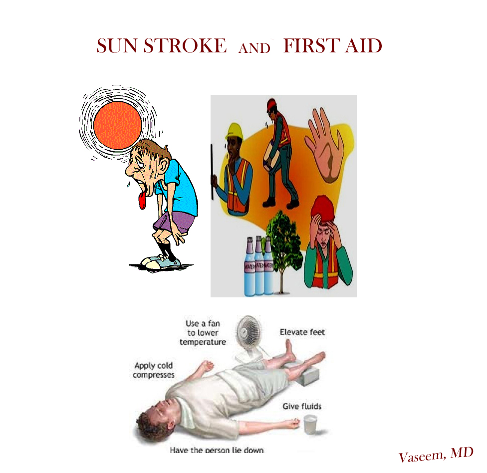
Q13 Could dehydration become so severe that one has to break the fast?

Yes. Harmful levels of water loss could occur if the person was poorly hydrated

before commencing the fast, and/or made worse by activities during the day and

weather conditions. If one produces very little or no urine, feels disorientated

and confused, or faints due to dehydration, the fast should be broken in order to

re-hydrate oneself.

Islam does not require that one harms him or herself in fulfilling the fast. If a fast is

broken, it will need to be compensated for by fasting at a later date.

Q14 Can I fast whilst I have dialysis?

Peritoneal dialysis requires the daily usage of fluid bags in the abdomen, and such

patients are advised not to fast (please refer to fidyah* below). Hemodialysis is

performed about 3 times a week, and results in significant shifts of fluids and salts

within the body. Such patients are also advised not to fast (please refer to fidyah*

below).

*Fidyah: is a method of compensation in Islam for a missed act of worship that must

be otherwise fulfi lled. If one is unable to fulfi ll a missed fast, for example due to an

ongoing illness should feed a hungry person (two meals per day) if he or she is able

to. Please consult an Islamic scholar for further details.

I wish you all to get the best out of Ramadhan.

Acknowledgements:

Dr Razeen Mahroof, BM MRCP(UK) FRCA, Anaesthetist, Oxford

Dr Rizwan Syed, BM DRCLG, General Practitioner, Birmingham

Dr. Ahmed El-Sharkawy, BM MRCP(UK), Specialist Registrar in

Gastroenterology

Tehseen Hasan, BSc (Hons), State Registered Dietitian (SRD), Birmingham

Henrietta Szovati, Researcher, Communities in Action

Sahra Ahmed MPharm, Pharmacist, Manchester

Tagged , , , , , , , , , , , , , , , , , ,

Can anybody understand me?

Image

I am a living, breathing and understanding human being. I feel light as all others do, I feel air as all others do, I feel pain as all others do and I wish I too could live normal life as all others do.

I am being misunderstood by everyone right from the pregnancy, birth and after delivery including my Mom and physicians. I suffer, not because I want to, but because of my disabilities and inability to explain what I am going through.

Many things I want to do, I want to say, I want to show but My cortical area of the brain does not let me to fulfill the task.

When I want to talk – people around me think, I am drooling.

When I want to walk – people around me think, I am nervous.

When I want to play – people around me think, I am mentally retarded with no proper movements.

I don’t want to laugh, but it is the cortical region which makes me laugh out of no reason.

I don’t want to limp, but it is the cortical region which causes uncontrollable spasms in my muscles.

I don’t want to be sick, but my body and brain are incompatible towards new infections and diseases and my immune system never got a chance to grow.

I want to be self sufficient but it is the uncoordinated nervous system which doesn’t let me to.

I see everyday normal people around me and I see how granted they take all those senses and movements for which I spend enough energy to get one.

I don’t want to be looked after as invalid or handicap, but help me very ‘little’ to define and learn the movements through continuous physical exercises and therapy.

A speech therapist to teach me effective speaking.

Last but not least a strong person – who can influence me while I work hard to get better.

Hope someday people will understand me. (A child with cerebral palsy)

Tagged , , , , , , ,

Cor Pulmonale – a student guide

COR PULMONALE

 It is the alteration of right ventricular structure or function that is due to pulmonary hypertension caused by diseases affecting the lung or its vasculature, NOT  LEFT HEART DIEASE causing right-heart-dis or congenital heart malformations.

Image

Image

NOTE RIGHT VENTRICULAR MUSCLE HYPERTROPHY*

PATHOPHYSIOLOGY of cor pulmonale:

Image

dyspnea on exertion, fatigue, lethargy, chest pain, syncope with exertion, pitting peripheral edema, passive hepatic congestion may lead to complaints such as anorexia and right upper quadrant discomfort.

Image

Auscultation of the heart also may reveal a systolic ejection murmur and, in more severe disease, a diastolic pulmonary regurgitation murmur.

b) Right ventricular hypertrophy is characterized by a prominent A wave in the jugular venous pulse, associated with a right-sided fourth heart sound and either a left parasternal heave or a downward subxiphoid thrust.

c) Right ventricular failure leads to systemic venous hypertension. This can produce a variety of findings, such as elevated jugular venous pressure with a prominent V wave, a right ventricular third heart sound, and a high-pitched tricuspid regurgitant murmur

Image

EVALUATION: Chest radiography
•  Electrocardiography
•  Two dimensional and Doppler echocardiography (which can provide an indirect measurement of pulmonary artery pressure when tricuspid regurgitation is present)
•  Pulmonary function tests
•  Radionuclide ventriculography
•  Magnetic resonance imaging
•  Right heart catheterization
•  Lung biopsy

TREATMENT: 1.Long term oxygen therapy

–         Oxygen therapy relieves pulmonary vasoconstriction, thereby decreasing pulmonary vascular resistance; as a result, the right ventricle increases stroke volume and cardiac output. Renal vasoconstriction also may be relieved, resulting in an increase in urinary sodium excretion.

Oxygen therapy improves arterial oxygen content, providing enhanced delivery to the heart, brain, and other vital organs.

Image           Image

2. Diuretics — If right ventricular filling volume is markedly elevated, diuretic therapy might improve the function of both right and left ventricles.

3. Vasodilators — Several vasodilator agents (including hydralazine, nitrates, nifedipineverapamil, and ACE inhibitors) have been utilized in an attempt to ameliorate pulmonary hypertension.

4. Theophylline and the sympathomimetic amines (terbutaline, etc) may have salutary effects not related to bronchodilation. Specifically, these agents may:
•  Improve myocardial contractility
•  Provide some degree of pulmonary vasodilation
•  Enhance diaphragm endurance

5. Phlebotomy — In patients with severe polycythemia (hematocrit above 55 percent).

Tagged , , , , , , , , , , , , , , , , , , , ,

The Cycle of Violence, Power and Control

the content (abuse of young girls) is so true not only for developed countries but also for developing and under-developed nations (world-wide)……
We need to look at basic cause for this behavior (economic crisis/instability?!?!? or no proper parenting?!?!?)

Licensed Mental Health Counselor

ImageWorking as a counselor in a high school, I am surprised at the amount of abuse many young girls I work with have gone through. Not to mention the sexual, physical and psychological abuse many of them went through growing up, but how much of that has affected them now as teenagers.

A surprising amount of young ladies in high school, and perhaps even in middle school are involved in physically abusive relationships. Having dealt with many of these young ladies, I’ve recognized that many of them believe that if a guy doesn’t hit or get physically rough with them, then “he doesn’t really love me”. This may not make any sense to most people, but a lot of these young ladies have grown up in homes where the people who “love” them, especially the men in their lives, are often the same people who abuse them, so many of…

View original post 590 more words